Image Acquisition and Processing
Pixel is the smallest discrete element of a digital image, representing a single point of intensity or colour. In radiology, each pixel corresponds to a specific location on the detector surface and records the amount of radiation that has …
Pixel is the smallest discrete element of a digital image, representing a single point of intensity or colour. In radiology, each pixel corresponds to a specific location on the detector surface and records the amount of radiation that has been absorbed. The size of a pixel, often expressed in millimetres, directly influences the spatial resolution of the image; smaller pixels can depict finer anatomical detail but may increase the amount of data that must be processed and stored. For example, a digital radiography detector with a pixel size of 0.1 Mm will resolve structures that are approximately 0.2 Mm apart, whereas a detector with a 0.2 Mm pixel size may miss those same structures.
Voxel extends the concept of a pixel into three dimensions. It is a cubic volume element that represents a unit of data in a volumetric dataset such as a computed tomography (CT) or magnetic resonance imaging (MRI) scan. The dimensions of a voxel are defined by the in‑plane pixel size and the slice thickness. If a CT scan has a pixel size of 0.5 Mm and a slice thickness of 1 mm, each voxel measures 0.5 Mm × 0.5 Mm × 1 mm. Understanding voxel geometry is essential for accurate measurement of organ volumes and for applying quantitative analysis techniques such as attenuation coefficient calculation.
Matrix refers to the two‑dimensional array of pixels that forms an image. In digital radiography, a common matrix size is 2048 × 2048, which yields over four million pixels per image. Larger matrices provide higher spatial resolution but increase the demand on storage capacity and network bandwidth when images are transferred to a picture archiving and communication system (PACS). In CT, the matrix is typically 512 × 512, although high‑resolution protocols may use 1024 × 1024 matrices.
Bit depth denotes the number of binary digits used to represent the intensity value of each pixel. An 8‑bit image can encode 256 shades of grey, while a 12‑bit image can encode 4096 shades. Higher bit depth improves the ability to differentiate subtle differences in tissue attenuation, which is particularly important in low‑contrast modalities such as soft‑tissue CT or MRI. However, increasing bit depth also enlarges file sizes and may require more sophisticated processing algorithms to avoid data overflow.
Dynamic range is the ratio between the highest and lowest detectable signal levels. In radiography, a wide dynamic range enables the capture of both high‑density bone and low‑density soft tissue within the same exposure. Modern flat‑panel detectors typically have a dynamic range of 14–15 bits, allowing them to handle a wide span of exposure levels without saturation. A limited dynamic range can lead to loss of detail in either the shadows (under‑exposure) or highlights (over‑exposure).
Signal‑to‑noise ratio (SNR) quantifies the proportion of true signal to random variations (noise) present in the image. SNR is calculated by dividing the mean signal intensity by the standard deviation of the background noise. Higher SNR values correspond to clearer images with less graininess. For example, a chest radiograph acquired at 70 kVp and 10 mAs may produce an SNR of 30, whereas increasing the mAs to 20 mAs can raise the SNR to approximately 42, improving diagnostic confidence but also raising patient dose.
Contrast‑to‑noise ratio (CNR) assesses the ability to distinguish a lesion from adjacent tissue, taking into account both contrast and noise. CNR is calculated as the difference in signal between the lesion and background divided by the noise level. A higher CNR indicates that subtle lesions, such as early‑stage tumours, are more likely to be detected. Optimising CNR often involves adjusting parameters such as tube voltage, filtration, and reconstruction algorithms.
Modulation transfer function (MTF) describes how an imaging system reproduces contrast at different spatial frequencies. It is a frequency‑domain representation of the system’s ability to resolve detail. An MTF curve that remains high at 5 lp/mm (line pairs per millimetre) indicates excellent resolution, whereas a rapid drop-off suggests poorer performance. MTF is measured using specialised phantoms and is a key metric in quality assurance programmes for digital radiography and CT.
Point spread function (PSF) characterises the response of an imaging system to a point source of radiation. In practice, the PSF is approximated by imaging a very small object, such as a metal bead, and analysing the resulting blur pattern. The width of the PSF, often expressed as full width at half maximum (FWHM), directly influences spatial resolution. A narrow PSF (small FWHM) indicates minimal blurring, which is desirable for high‑resolution imaging.
Line spread function (LSF) is similar to the PSF but derived from a line source instead of a point source. It is useful for evaluating systems that produce linear features, such as the edges of a radiographic grid. The LSF is also used to calculate the MTF through Fourier transformation.
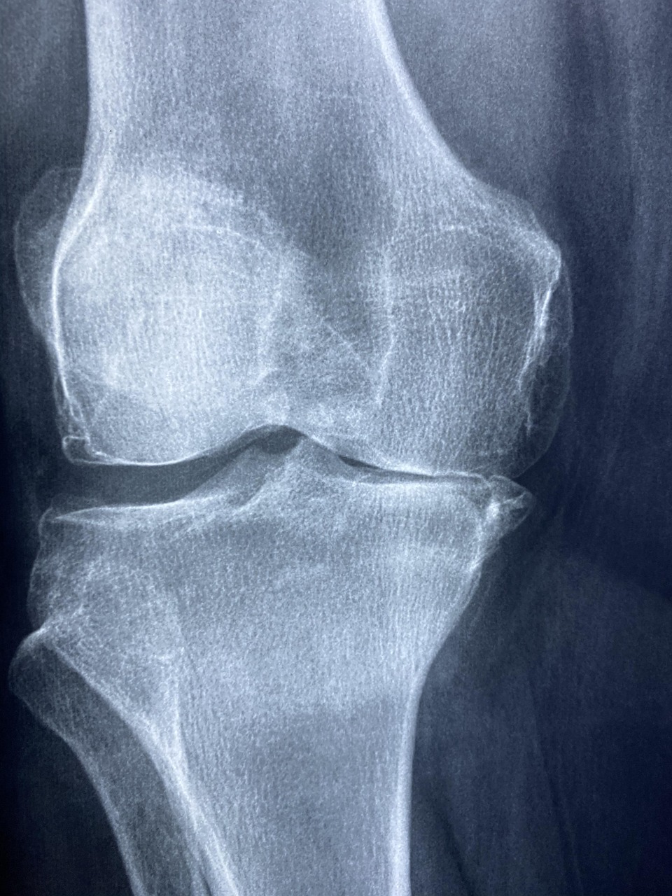
Spatial resolution is the ability of an imaging system to distinguish small objects that are close together. It is commonly expressed in line pairs per millimetre (lp/mm) or as the minimum resolvable distance in millimetres. Spatial resolution is affected by pixel size, detector blur, focal spot size, and patient motion. In mammography, spatial resolution requirements are stringent; a system must resolve at least 5 lp/mm to detect microcalcifications.
Temporal resolution refers to the capability of a system to capture rapidly changing events. In fluoroscopy, high temporal resolution (e.G., 30 Frames per second) is essential for visualising dynamic cardiac motion. Temporal resolution is limited by detector readout speed and processing latency. In MRI, temporal resolution is expressed as the acquisition time per image set; faster sequences reduce motion artefacts but may compromise signal strength.
Exposure denotes the amount of ionising radiation delivered to the detector or patient. It is measured in units of milliampere‑seconds (mAs) for X‑ray tubes, reflecting the product of tube current (mA) and exposure time (s). Adjusting exposure influences image brightness and noise; higher exposure reduces noise but increases dose, while lower exposure reduces dose but may increase graininess.
Dose is the amount of radiation absorbed by the patient’s tissues, quantified as the absorbed dose (gray, Gy) or effective dose (millisievert, mSv). Dose management is a central concern in radiology quality assurance. For instance, a standard chest X‑ray delivers approximately 0.1 MSv, whereas a CT abdomen‑pelvis study may deliver 8–10 mSv. Strategies such as automatic exposure control (AEC) and dose‑modulation algorithms aim to keep dose as low as reasonably achievable (ALARA) while preserving diagnostic image quality.
kVp (kilovolt peak) defines the maximum voltage applied across the X‑ray tube, determining the energy spectrum of the emitted photons. Higher kVp produces more penetrating beams, reducing contrast but also decreasing patient dose because fewer photons are absorbed in the body. Typical chest radiographs use 110–130 kVp, whereas bone studies may require 60–80 kVp to enhance contrast between bone and soft tissue.
mAs (milliampere‑seconds) controls the quantity of X‑ray photons generated. Increasing mAs raises the number of photons, improving SNR but also raising dose. Modern radiography units often employ automatic mAs selection based on patient size and desired image quality, thereby standardising exposure across diverse patient populations.
Automatic exposure control (AEC) is a feedback system that measures the amount of radiation reaching the detector and terminates the exposure once a pre‑set detector exposure level is achieved. AEC improves consistency between examinations and reduces the risk of over‑ or under‑exposure. In practice, AEC sensors are positioned behind the patient, and the system adjusts exposure time and tube current in real time.
Filtration involves placing metal plates (commonly aluminium or copper) in the X‑ray beam path to absorb low‑energy photons that would otherwise increase patient dose without contributing to image formation. The effective filtration is expressed in millimetres of aluminium equivalent (mm Al). For example, a 2.5 Mm Al filter reduces the proportion of low‑energy photons, thereby improving beam quality and reducing skin dose.
Scatter refers to photons that deviate from their original trajectory after interacting with matter. Scatter degrades image contrast by adding unwanted background signal. Anti‑scatter grids, composed of lead strips, are used to absorb scattered photons while allowing primary photons to reach the detector. The grid ratio (e.G., 6:1) Indicates the height-to-spacing of the lead strips; higher ratios provide better scatter rejection but increase exposure requirements.
Beam hardening is a phenomenon where low‑energy photons are preferentially absorbed as the beam passes through dense material, causing the effective beam energy to increase (harden). In CT, beam hardening leads to cupping artefacts—central regions appear darker than peripheral regions. Correction algorithms, such as linearisation or iterative reconstruction, are employed to mitigate these artefacts.
Partial volume effect occurs when a voxel contains a mixture of tissues with different attenuation coefficients, resulting in an averaged signal that may not accurately represent any individual component. This effect can obscure small lesions, especially when slice thickness exceeds the size of the structure. Reducing slice thickness or applying high‑resolution reconstructions can lessen partial volume artefacts.
Reconstruction is the computational process of converting raw projection data into a cross‑sectional image. In CT, filtered back‑projection (FBP) has been the traditional method; it applies a convolution filter to each projection before back‑projecting it into image space. Modern CT systems increasingly use iterative reconstruction (IR) techniques, which model the physics of data acquisition and noise statistics to produce images with reduced noise at lower dose levels.
Filtered back‑projection (FBP) relies on a mathematical filter—commonly a Ram‑Lak or Hann filter—to enhance high‑frequency components before back‑projecting the data. While computationally efficient, FBP can amplify noise, especially at low dose, leading to grainy images. Consequently, FBP is gradually being supplanted by more sophisticated iterative methods in dose‑sensitive applications.
Iterative reconstruction (IR) algorithms iteratively refine an image estimate by comparing simulated projections of the current estimate with the actual measured data. Each iteration reduces the discrepancy, allowing for noise suppression and improved contrast resolution. Popular IR approaches include statistical IR (e.G., ASIR, SAFIRE) and model‑based IR (e.G., MBIR). IR can achieve dose reductions of 30–50 % while maintaining diagnostic quality, though it may introduce a “plastic” appearance that requires radiologists to adapt.
Windowing and leveling are post‑processing adjustments that map the wide range of pixel values onto the display’s limited grayscale. The window width defines the range of intensities displayed, while the window level sets the centre of that range. For a chest CT, a window width of 1500 HU and level of –500 HU highlights lung parenchyma; a bone window might use a width of 2000 HU and level of 500 HU to visualise cortical bone. Proper windowing is essential for accurate interpretation and is often taught as a fundamental skill in radiology training.
Histogram equalisation is an image‑enhancement technique that redistributes pixel intensity values to achieve a uniform histogram. This process can improve contrast in under‑exposed images but may also amplify noise. In practice, radiology software provides adaptive histogram equalisation, which applies the technique locally to preserve details while enhancing overall visibility.
Noise reduction filters, such as Gaussian smoothing, median filtering, or non‑local means, are employed to suppress random variations without overly blurring edges. Gaussian filters apply a weighted average based on distance from the central pixel, reducing high‑frequency noise. Median filters replace each pixel with the median value of its neighbourhood, effectively removing impulse noise (salt‑and‑pepper artefacts). Non‑local means algorithms compare patches across the entire image to preserve texture while reducing noise, albeit at higher computational cost.
Edge detection algorithms, such as the Sobel or Canny operators, identify boundaries between regions of differing intensity. Edge detection is frequently used in automated segmentation pipelines to delineate organ contours or tumour margins. For instance, a Canny edge detector can isolate the sharp transition between a lung nodule and surrounding aerated lung, facilitating volumetric measurement.
Segmentation divides an image into meaningful regions based on intensity, texture, or anatomical criteria. Manual segmentation relies on radiologist input, while semi‑automated methods combine thresholding with region‑growing or active‑contour models (snakes). Fully automated deep‑learning approaches, such as convolutional neural networks (CNNs), have demonstrated high accuracy in segmenting structures like the liver, brain ventricles, and prostate, but they require extensive training datasets and rigorous validation to ensure reliability.
Artefact is any feature in an image that does not represent true anatomy, often arising from technical limitations or patient factors. Common artefacts include motion blur, metal streaks, beam‑hardening cupping, and aliasing. Recognising artefacts is vital for accurate diagnosis; for example, a metal prosthesis can produce streak artefacts that obscure adjacent bone, prompting the use of metal‑artefact reduction (MAR) algorithms.
Aliasing occurs when the sampling frequency (pixel size) is insufficient to capture high‑frequency information, leading to false patterns or moiré effects. In digital radiography, aliasing can manifest as a “checkerboard” appearance when the detector’s pixel pitch does not align with the X‑ray focal spot geometry. Anti‑aliasing filters and appropriate sampling strategies are employed to minimise this effect.
Motion artefact results from patient movement during image acquisition. In CT, patient motion can cause blurring or duplication of structures, especially in the thorax where cardiac and respiratory motion are pronounced. Strategies to reduce motion artefacts include breath‑hold instructions, faster rotation speeds (e.G., 0.28 S per rotation), and prospective gating synchronized with the cardiac cycle.
Gating synchronises image acquisition with a physiological signal, typically the ECG for cardiac imaging. Prospective gating acquires data only during a predefined phase of the cardiac cycle (e.G., Diastole), reducing radiation exposure and motion artefacts. Retrospective gating records data throughout the cycle and reconstructs images at multiple phases, enabling functional assessment but at higher dose.
Phantom is a test object designed to mimic the radiographic properties of human tissue. Phantoms are used for routine quality control, calibration, and performance verification of imaging systems. Common phantoms include the Leeds TOR test object for evaluating spatial resolution and contrast, the CT head phantom for uniformity and low‑contrast detectability, and the ACR mammography phantom for assessing microcalcification detection.
Calibration aligns the output of an imaging system with known standards. In radiography, detector calibration ensures that the pixel values correspond accurately to the incident radiation dose. Regular calibration checks, performed weekly or monthly depending on departmental policy, help maintain consistent image quality and prevent drift in detector response.
Quality control (QC) programmes encompass a series of systematic tests designed to verify that imaging equipment operates within predefined specifications. QC activities include daily exposure checks, weekly uniformity assessments, monthly spatial resolution measurements, and annual comprehensive evaluations. Documentation of QC results is essential for regulatory compliance and for tracking performance trends over time.
DICOM (Digital Imaging and Communications in Medicine) is the universal standard for handling, storing, and transmitting medical images. DICOM defines file formats, network protocols, and metadata structures. Understanding DICOM tags—such as Patient ID, Study Instance UID, and Image Position Patient—is crucial for ensuring correct image identification and for integrating with PACS and radiology information systems (RIS).
PACS (Picture Archiving and Communication System) stores and provides access to digital images across the radiology department. PACS architecture typically includes image acquisition servers, storage arrays, and viewer workstations. Efficient PACS operation relies on proper network bandwidth, data redundancy, and regular backup procedures. Radiologists must be familiar with PACS navigation, image retrieval, and annotation tools to optimise workflow.
Radiation safety encompasses measures to protect patients, staff, and the public from unnecessary exposure. Key concepts include ALARA, shielding, dose monitoring, and the use of protective equipment such as lead aprons and thyroid collars. In the context of image acquisition, radiation safety is balanced against the need for sufficient image quality to answer the clinical question.
Contrast agents enhance the visibility of vascular structures, organs, or lesions by altering the attenuation properties of the targeted area. Iodinated contrast is used in CT angiography, while gadolinium‑based agents are employed in MRI. Understanding the timing of contrast injection, bolus tracking, and appropriate dosing is essential for acquiring diagnostic images and for preventing adverse reactions.
Bolus tracking monitors the arrival of contrast material in a region of interest using real‑time attenuation measurements. Once a predefined enhancement threshold is reached, the scanner initiates image acquisition. Bolus tracking improves timing accuracy for dynamic studies such as CT pulmonary angiography, reducing the risk of suboptimal opacification.
Dynamic imaging captures a series of images over time to evaluate physiological processes. In fluoroscopy, dynamic imaging is used for interventional procedures, while in MRI, dynamic contrast‑enhanced (DCE) sequences assess tumour perfusion. Temporal resolution, contrast injection rate, and post‑processing algorithms (e.G., Kinetic modelling) are critical factors in producing meaningful dynamic data.
Multi‑detector CT (MDCT) employs multiple rows of detector elements, enabling rapid acquisition of volumetric data with thin slices. MDCT systems with 64, 128, or 256 detector rows can cover large anatomical volumes in a single rotation, reducing motion artefacts and allowing for high‑resolution reconstructions. However, the increased data throughput demands robust processing hardware and efficient storage solutions.
Dual‑energy CT (DECT) acquires data at two distinct tube voltages (e.G., 80 KVp and 140 kVp) either sequentially or simultaneously using dual‑source scanners. DECT enables material decomposition, allowing differentiation of iodine, calcium, and uric acid, as well as virtual non‑contrast imaging. This capability can reduce the need for additional scans, thereby lowering overall radiation dose.
Magnetic resonance imaging (MRI) uses strong magnetic fields and radiofrequency pulses to generate images based on the nuclear magnetic resonance of hydrogen nuclei. Key parameters influencing image quality include field strength (1.5 T versus 3 T), echo time (TE), repetition time (TR), and flip angle. MRI does not involve ionising radiation, making it preferable for repeated follow‑up examinations, but it is sensitive to motion and requires careful patient screening for metallic implants.
Spin‑echo and gradient‑echo are fundamental pulse sequences in MRI. Spin‑echo sequences provide high‑quality T1‑ or T2‑weighted images with excellent contrast but longer acquisition times. Gradient‑echo sequences are faster and enable imaging of susceptibility effects (e.G., Blood products) but are more prone to artefacts. Selecting the appropriate sequence balances diagnostic requirements against scan duration and patient comfort.
Diffusion‑weighted imaging (DWI) measures the random motion of water molecules within tissue, providing information about cellular density and membrane integrity. Apparent diffusion coefficient (ADC) maps derived from DWI are valuable in oncology for tumour characterisation and in stroke imaging for early detection of ischemic changes. High b‑values increase diffusion weighting but reduce signal‑to‑noise ratio, necessitating optimisation of acquisition parameters.
Functional MRI (fMRI) detects changes in blood oxygenation level dependent (BOLD) signals during neural activation. FMRI is employed in pre‑surgical planning to map eloquent cortex. Temporal resolution (typically 2–3 s per volume) and spatial resolution (3–4 mm voxels) are trade‑offs; higher temporal resolution improves detection of rapid neural events, while higher spatial resolution enhances localisation accuracy.
Positron emission tomography (PET) visualises metabolic activity by detecting pairs of gamma photons emitted from positron‑emitting radiotracers (e.G., 18F‑FDG). PET images are inherently low‑resolution and noisy; reconstruction algorithms such as ordered‑subset expectation maximisation (OSEM) improve image quality. PET is frequently combined with CT (PET/CT) to provide anatomical localisation, and the fusion of PET with MRI (PET/MRI) offers superior soft‑tissue contrast.
SPECT (Single Photon Emission Computed Tomography) uses gamma‑emitting isotopes (e.G., 99MTc) and rotating gamma cameras to produce three‑dimensional images. SPECT resolution is lower than PET, but it remains valuable for functional cardiac imaging and bone scintigraphy. Attenuation correction and scatter correction are essential post‑processing steps to enhance quantitative accuracy.
Attenuation correction compensates for the reduction in photon intensity as it traverses the body. In PET, attenuation maps derived from CT data are applied to correct the raw sinograms, improving quantitative fidelity. In SPECT, attenuation correction may be performed using transmission scans or model‑based approaches. Failure to apply appropriate attenuation correction can lead to underestimation of tracer uptake in deep tissues.
Scatter correction removes the contribution of scattered photons that degrade image contrast. In PET, scatter correction algorithms estimate the scattered component from the measured data and subtract it before reconstruction. In CT, scatter correction is implemented through hardware (e.G., Anti‑scatter grids) and software (e.G., Iterative correction). Accurate scatter correction enhances low‑contrast detectability and quantitative reliability.
Metal‑artefact reduction (MAR) algorithms address streak artefacts caused by high‑density implants. MAR techniques typically involve segmenting the metal, forward projecting its influence, and iteratively correcting the raw data. While MAR can restore visibility of adjacent structures, it may introduce new artefacts or alter attenuation values, requiring radiologists to interpret the corrected images with caution.
Image registration aligns images from different modalities, time points, or patient positions into a common coordinate system. Rigid registration accounts for translation and rotation, while deformable registration handles non‑linear anatomical changes. Accurate registration is essential for multimodality fusion (e.G., PET/CT), longitudinal studies, and image‑guided interventions.
Image fusion combines information from two or more imaging modalities to create a composite image that leverages the strengths of each. For example, fusing a high‑resolution CT angiogram with a PET metabolic map can delineate the extent of a tumour’s vascular supply. Fusion requires precise registration and may involve colour‑coding to differentiate the contributing datasets.
Radiomics extracts quantitative features (e.G., Texture, shape, intensity) from medical images for predictive modelling. Radiomic pipelines involve segmentation, feature extraction, dimensionality reduction, and statistical or machine learning analysis. Radiomics holds promise for personalised medicine, but challenges include reproducibility across scanners, standardisation of acquisition parameters, and the need for large, annotated datasets.
Deep learning in medical imaging utilizes neural networks with many layers to perform tasks such as classification, segmentation, and anomaly detection. Convolutional neural networks (CNNs) have become the backbone of many diagnostic assistance tools, automatically identifying pathologies like lung nodules or intracranial haemorrhage. Deployment of deep‑learning models requires rigorous validation, bias assessment, and integration with existing workflow to ensure patient safety.
Standardisation of acquisition protocols is vital for consistent image quality and reliable quantitative analysis. Protocol standardisation involves defining parameters such as kVp, mAs, slice thickness, reconstruction kernel, and contrast timing. In multi‑centre studies, adherence to a common protocol reduces variability and facilitates pooled data analysis.
Reconstruction kernel (or filter) determines how raw data are processed to produce the final image. Soft‑tissue kernels (e.G., “Standard”) preserve low‑frequency information, yielding smoother images suitable for organ assessment. High‑frequency kernels (e.G., “Bone”) enhance edge definition, improving the visibility of fine bony structures but increasing noise. Selecting an appropriate kernel is a trade‑off between resolution and noise.
Slice thickness influences both spatial resolution and dose. Thin slices (e.G., 0.5 Mm) improve detection of small lesions and enable high‑resolution multiplanar reconstructions, but they increase image noise and data volume. Thick slices (e.G., 5 Mm) reduce noise and dose but may obscure fine detail. Many protocols acquire thin slices for reconstruction while storing thicker averages for routine viewing.
Pitch in helical CT denotes the table feed per rotation divided by the total width of the X‑ray beam. A pitch of 1.0 Means the table moves a distance equal to the beam width during one rotation. Lower pitch values (e.G., 0.5) Increase overlap, improving image quality at the cost of higher dose, whereas higher pitches (e.G., 1.5) Reduce dose but may introduce gaps and reduce resolution. Optimising pitch is essential for balancing dose and diagnostic yield.
Automatic tube current modulation (ATCM) adjusts the tube current in real time based on patient attenuation, delivering higher current for denser regions and lower current for less attenuating areas. ATCM reduces overall dose while maintaining image quality. Modern systems provide angular, longitudinal, and combined modulation modes, each with specific settings that must be calibrated during quality assurance checks.
Image compression reduces file size for storage and transmission. Lossless compression (e.G., JPEG‑2000 lossless) preserves all original data, making it suitable for archival and quantitative analysis. Lossy compression (e.G., JPEG) discards some information, potentially affecting diagnostic quality if compression ratios are excessive. Regulatory guidelines often specify maximum allowable compression levels for diagnostic images.
Viewer workstation hardware and software must meet performance criteria to support high‑resolution image display, rapid manipulation (zoom, pan, rotate), and advanced processing (e.G., 3‑D rendering). Calibration of display monitors includes verification of luminance, colour accuracy, and uniformity according to standards such as DICOM Part 14. Regular monitor quality assurance ensures that subtle differences in grayscale are faithfully reproduced.
Radiation dose monitoring systems track cumulative exposure for individual patients and staff. Dose‑tracking software aggregates data from DICOM Radiation Dose Structured Reports (RDSR) and provides alerts when predefined thresholds are approached. Implementing dose monitoring helps identify outlier examinations, guides protocol optimisation, and supports compliance with national dose‑reporting requirements.
Shielding devices, such as lead aprons, thyroid collars, and gonadal shields, protect radiosensitive tissues from scatter radiation. In interventional radiology, ceiling‑mounted lead shields and table‑top shields are employed to reduce staff exposure. Proper positioning and regular inspection of shielding equipment are essential to maintain effectiveness.
Quality assurance (QA) audit involves periodic review of equipment performance, protocol adherence, and documentation. Audits may be internal (departmental) or external (regulatory bodies). An effective QA audit includes trend analysis of QC metrics, identification of deviations, corrective action plans, and documentation of outcomes. Audits support continuous improvement and help maintain accreditation status.
Regulatory standards governing radiology quality assurance in the United Kingdom include the Ionising Radiation (Medical Exposure) Regulations (IRMER) and the British Institute of Radiology (BIR) guidelines. These documents outline legal responsibilities for dose optimisation, equipment testing frequencies, and record‑keeping. Compliance with these standards is mandatory for licencing and for participation in national audit programmes such as the Radiology Clinical Effectiveness Group (RCEG).
Clinical decision support (CDS) tools integrate patient data with evidence‑based imaging guidelines (e.G., NICE criteria) to recommend appropriate imaging studies. CDS helps prevent unnecessary examinations, reducing radiation exposure and resource utilisation. Integration of CDS with radiology information systems enables real‑time alerts if a requested study falls outside recommended parameters.
Patient positioning influences image quality and dose distribution. Correct positioning reduces the need for repeat examinations and optimises the alignment of the anatomy with the detector. For example, in chest radiography, the patient should be centred on the detector with the central ray angled 10–15° cephalad to compensate for thoracic curvature, thereby improving lung field coverage and reducing magnification of the heart.
Collimation shapes the X‑ray beam to match the area of clinical interest, limiting exposure to surrounding tissues. Proper collimation reduces scatter, improves contrast, and lowers patient dose. In digital radiography, automatic collimation sensors can adjust the beam size dynamically, but radiographers must verify that the selected field matches the clinical requirement.
Grid alignment ensures that the anti‑scatter grid’s lead strips are parallel to the detector plane. Misalignment causes grid cut‑off, resulting in a loss of primary photons and increased image noise. Routine grid checks involve visual inspection of the grid’s orientation and verification of uniform exposure across the detector.
Exposure index (EI) is a parameter reported by many digital detectors that reflects the amount of radiation exposure received by the detector. EI values are compared against target ranges defined by the manufacturer and the relevant standards (e.G., IEC 62494‑1). Deviations from the target EI indicate under‑ or over‑exposure, prompting adjustments to technique factors.
Noise index is a user‑defined target value for image noise, used in automatic exposure control algorithms. The system automatically modulates tube current to achieve the specified noise index, thereby standardising image quality across patients of varying size. Selecting an appropriate noise index balances diagnostic adequacy with dose reduction.
Contrast enhancement techniques, such as high‑pass filtering or adaptive histogram equalisation, increase the visibility of low‑contrast structures. While useful for visual interpretation, excessive enhancement can create artificial edges that may mislead radiologists. Therefore, contrast enhancement should be applied judiciously, and original raw data should be available for verification.
3‑D rendering techniques, including volume rendering and maximum intensity projection (MIP), transform stacked axial images into three‑dimensional visualisations. Volume rendering displays the entire dataset with varying opacity, useful for assessing complex vascular anatomy. MIP projects the highest attenuation values along each line of sight, highlighting contrast‑filled vessels in CT angiography. Mastery of these tools enhances the radiologist’s ability to interpret intricate anatomical relationships.
Virtual reality (VR) and augmented reality (AR) are emerging platforms for interactive image exploration. VR immerses the user in a fully simulated environment, allowing manipulation of volumetric data in three dimensions. AR overlays imaging data onto the patient’s body during procedures, aiding image‑guided interventions. Both technologies require high‑resolution displays, accurate registration, and robust software pipelines to be clinically useful.
Radiation dose metrics such as CT dose index volume (CTDIvol) and dose‑length product (DLP) provide estimates of the radiation output of a CT scan. CTDIvol reflects the dose per slice, while DLP incorporates the scan length, yielding a more comprehensive dose estimate. Converting DLP to effective dose using region‑specific conversion factors enables comparison with diagnostic reference levels (DRLs).
Diagnostic reference levels (DRLs) are benchmark dose values derived from national surveys and represent typical practice for standard examinations. Exceeding a DRL prompts review of technique factors, patient positioning, and equipment performance. DRLs are not dose limits; rather, they serve as tools for dose optimisation and quality improvement.
Image artefact reduction strategies include hardware solutions (e.G., Improved detector design, stronger shielding) and software techniques (e.G., Iterative reconstruction, MAR). Understanding the origin of each artefact guides the selection of the most effective mitigation approach. For instance, ringing artefacts caused by high‑frequency noise can be reduced by applying a low‑pass filter during reconstruction.
Patient dose tracking databases collect exposure information across multiple examinations, enabling longitudinal monitoring of cumulative dose. Integration with electronic health records allows clinicians to assess the risk‑benefit ratio of repeat imaging, especially for patients with chronic conditions requiring frequent surveillance (e.G., Oncology follow‑up).
Radiation‑induced skin injury is a rare but serious complication of high‑dose interventional procedures. Early signs include erythema and desquamation, progressing to ulceration if unrecognised. Preventative measures include limiting fluoroscopy time, using low‑dose protocols, and employing skin dose mapping software that visualises cumulative dose distribution on the patient’s anatomy.
Motion correction algorithms address patient movement during acquisition. In MRI, prospective motion correction uses navigator echoes to adjust gradients in real time, while retrospective correction applies image registration post‑acquisition. In CT, motion correction may involve data‑sorting based on cardiac phase or using deformation fields to align projection data before reconstruction.
Phantom studies replicate clinical scenarios to evaluate the performance of new acquisition techniques or reconstruction algorithms. For example, a low‑contrast phantom can assess the detectability of lesions at different dose levels, while a high‑resolution line‑pair phantom evaluates spatial resolution. Results from phantom studies inform protocol optimisation and equipment commissioning.
Equipment commissioning is the process of validating that a new imaging system meets manufacturer specifications and clinical requirements before routine use. Commissioning includes acceptance testing, baseline QC measurements, and training of staff. Documentation of commissioning results forms part of the quality management system and is required for regulatory compliance.
Training and competency programmes ensure that radiographers, technologists, and physicists possess the knowledge and skills to operate imaging equipment safely and effectively. Competency assessments may involve written exams, practical demonstrations, and periodic re‑certification. Ongoing education addresses emerging technologies such as AI‑driven image analysis and advanced dose‑reduction techniques.
Incident reporting provides a mechanism for documenting and analysing unexpected events, such as equipment failures, radiation over‑exposures, or image quality issues. A systematic incident reporting system enables root‑cause analysis, implementation of corrective actions, and prevention of recurrence. Reporting should be non‑punitive to encourage staff participation.
Data security and privacy are critical in handling medical images, which contain protected health information (PHI). Compliance with the General Data Protection Regulation (GDPR) mandates secure storage, controlled access, and encryption of image data. Radiology departments must implement robust access controls, audit trails, and regular security assessments to safeguard patient confidentiality.
Standard operating procedures (SOPs) codify the steps required for image acquisition, processing, and quality control. SOPs promote consistency, reduce variability, and provide a reference for training new staff. Examples of SOPs include “Daily detector warm‑up”, “Weekly uniformity check”, and “Protocol for contrast‑enhanced CT abdomen”.
Key takeaways
- The size of a pixel, often expressed in millimetres, directly influences the spatial resolution of the image; smaller pixels can depict finer anatomical detail but may increase the amount of data that must be processed and stored.
- Understanding voxel geometry is essential for accurate measurement of organ volumes and for applying quantitative analysis techniques such as attenuation coefficient calculation.
- Larger matrices provide higher spatial resolution but increase the demand on storage capacity and network bandwidth when images are transferred to a picture archiving and communication system (PACS).
- Higher bit depth improves the ability to differentiate subtle differences in tissue attenuation, which is particularly important in low‑contrast modalities such as soft‑tissue CT or MRI.
- Modern flat‑panel detectors typically have a dynamic range of 14–15 bits, allowing them to handle a wide span of exposure levels without saturation.
- For example, a chest radiograph acquired at 70 kVp and 10 mAs may produce an SNR of 30, whereas increasing the mAs to 20 mAs can raise the SNR to approximately 42, improving diagnostic confidence but also raising patient dose.
- Contrast‑to‑noise ratio (CNR) assesses the ability to distinguish a lesion from adjacent tissue, taking into account both contrast and noise.